


Detection
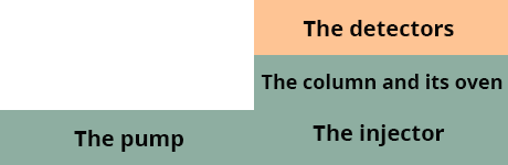
The different detectors
Many detectors can be used in HPLC. However, three detectors are mainly used today for detection in liquid chromatography:
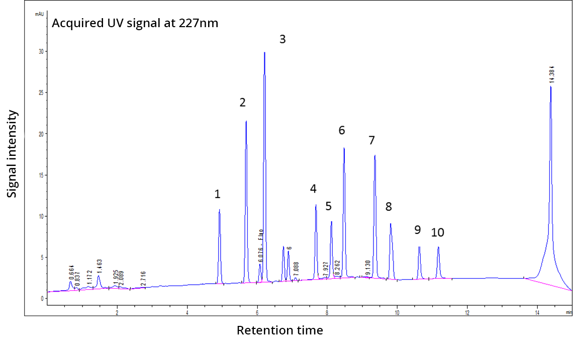
The chromatogram
The chromatogram is the 2D representation of the signal analyzed by chromatography. The two dimensions commonly found on a chromatogram are time and signal intensity (e.g. absorbance, luminescence, conductivity, m/z). In this way, the eluted compounds are represented by peaks on the chromatogram (visible in the figure below):
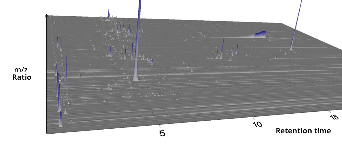
Some detectors are capable of collecting several signals simultaneously (several wavelengths or m/z) enabling three-dimensional chromatograms to be plotted. However, 3D chromatograms are not very often used in chromatography because the signal processing is complex and requires advanced mathematical tools.
Quantification
The quantification of the compounds analyzed in HPLC is based on the peaks visible on the chromatogram.
The useful parameter for quantification is either the area of the peak or its height. In general, quantification is preferred using peak area rather than peak height. The peak area is obtained using the integration software sold with chromatographic instruments.
On the chromatogram below you can see a peak that has been automatically integrated by the software. Despite the good quality of recent software, caution is advised for each peak integration as an incorrect integration can generate important errors in the quantification of the compounds.
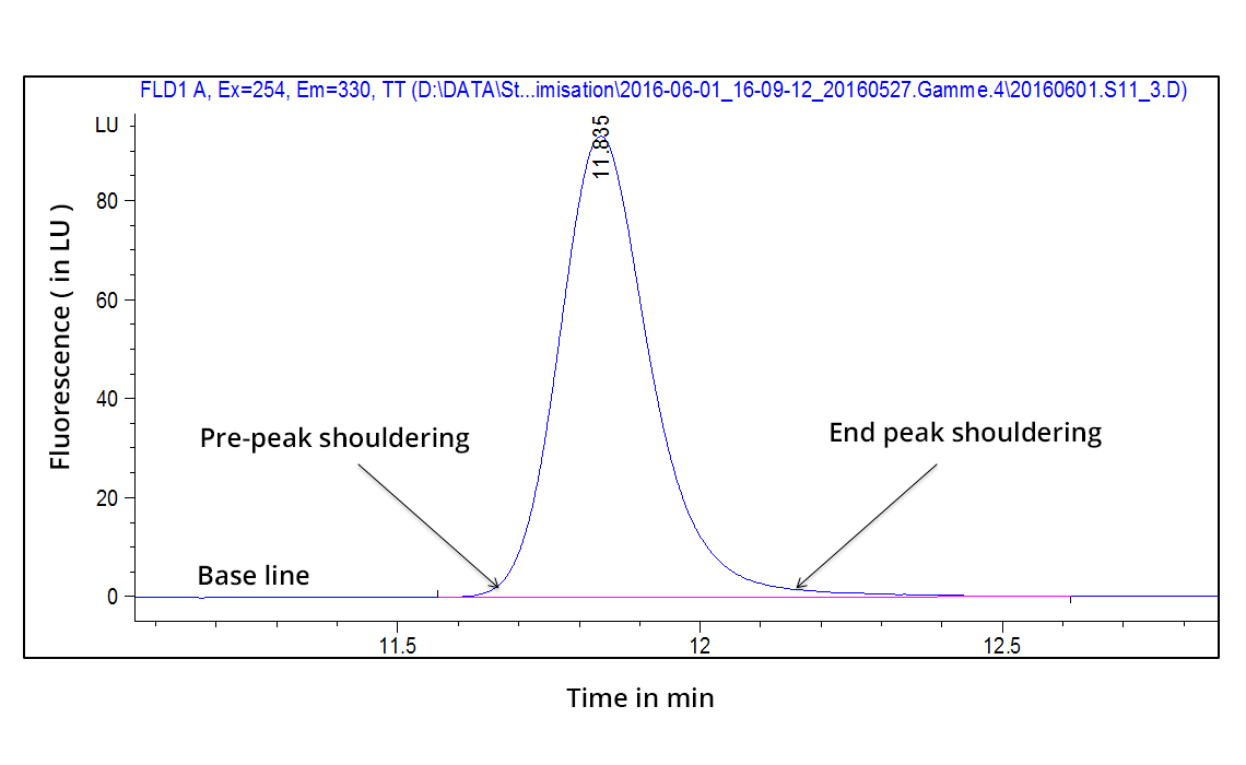
Chromatographic peak integration
For a quantification to be correct, some important points must be respected:
- The integration must start before the pre-peak shoulder
- The integration must finish after the end-peak shoulder
- The integration must not be too far from these two shoulders
- The integration line must be in line with the baseline of the chromatogram
If all these criteria are respected then the integration is correct and quantification can be done using the calibration method