Basics of UV-visible absorption spectroscopy
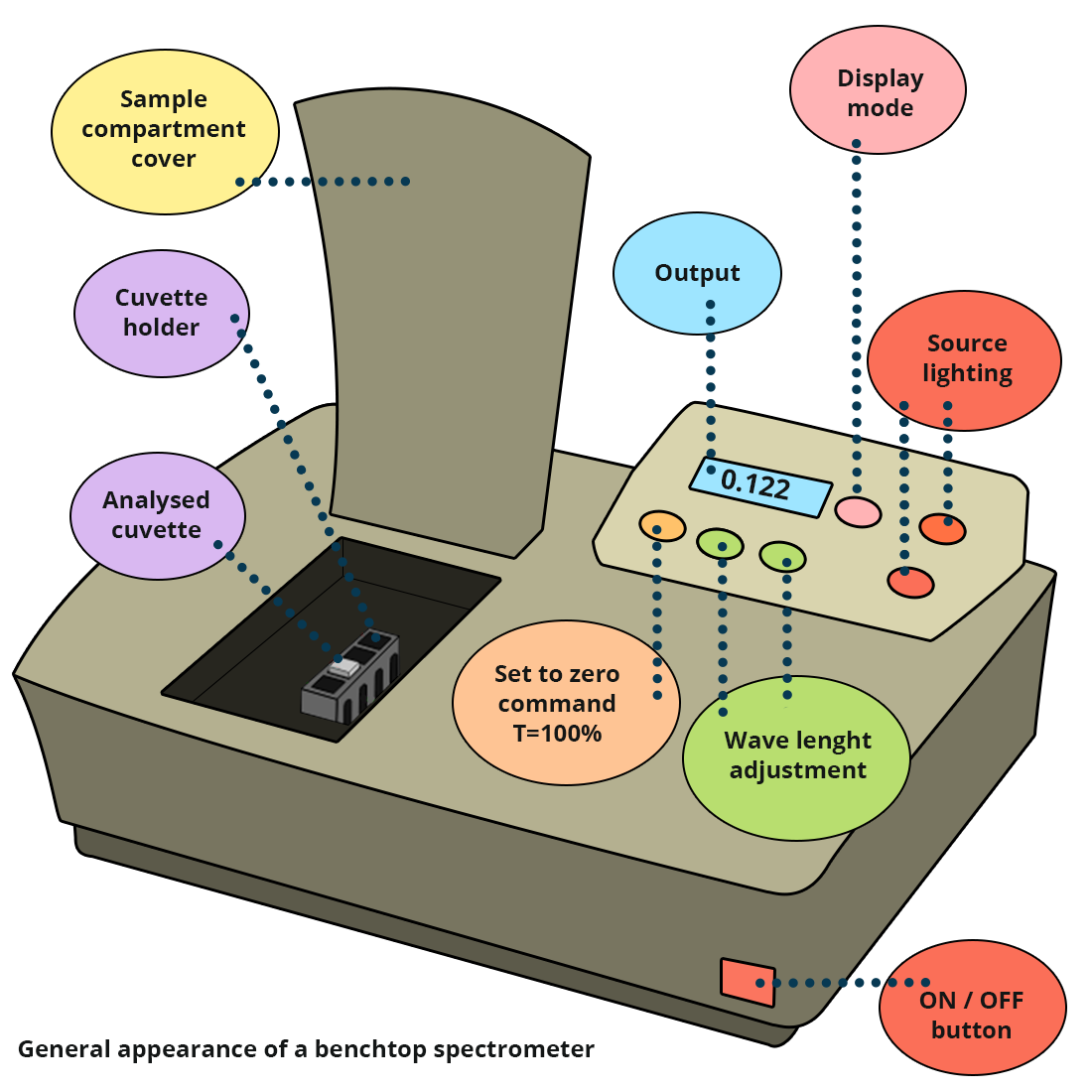
A spectrophotometer
A "single-beam" spectrophotometer is made up of:
- a polychromatic source
- a wavelength selector and eventually a source selector
- a sample compartment
- a detector
- a screen and/or a computer.


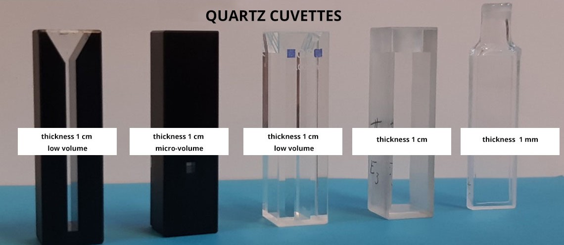
For liquids, cuvettes with a defined path length can be used. They can be in quartz, glass or plastic, for example, depending on the sample type.
 |
 |
 |
 |
 |
 |
 |
| 1 mm wide glass |
10 mm wide glass |
10 mm wide "quartz" |
50 mm wide glass |
10 mm wide glass, small volume |
10 mm wide glass, very small volume |
10 mm wide plastic, shrinked |
Powder samples are placed in a special sample holder (integrating sphere).

Schematic representation of a spectrophotometer
Click on each element to see the details

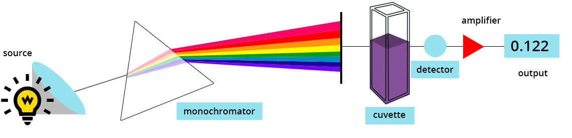
How does a spectrophotometer work?
"White" light is obtained from a polychromatic source, then a monochromator extracts the radiation at the wavelength requested by the user. Beam intensity is measured after it has passed through the sample in the cuvette. This intensity is compared to a reference value – the resulting value displayed by the instrument reflects this comparison. Thus, for a single-beam spectrophotometer, the reference spectrum is recorded with the solution (solvent or buffer) but without the sample. For a double-beam spectrophotometer, one cuvette contains the sample and the other one only contains the solution: the difference between both absorbances is then directly done by the system.